Dopamine D3 Receptor
|
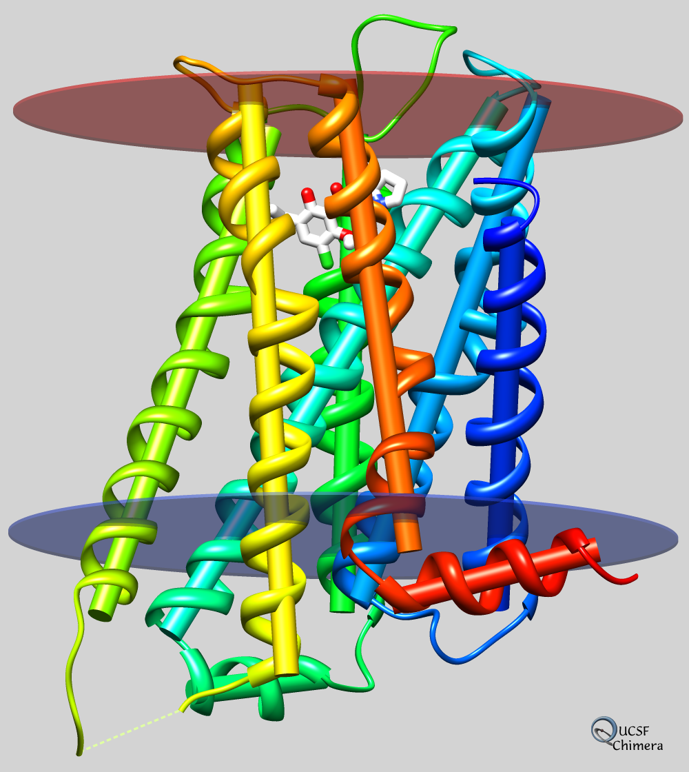
| larger image |
Dopamine activates several types of G protein-coupled receptors in the brain. The image shows the human D3 type dopamine receptor in complex with the antagonist eticlopride (Protein Data Bank entry 3pbl) as modeled into the membrane in the Orientations of Proteins in Membranes (OPM) database. The inner and outer boundaries of the membrane are indicated with transparent blue and red disks, respectively. The receptor is rainbow-color-coded from blue at the N-terminus to red at the C-terminus, and the axis of each helix is shown as a cylinder. Axes, planes, and centroids representing sets of atoms can be created with the command define or the Axes/Planes/Centroids tool, and these objects can be used in distance and angle measurements.
References:
Structure of the human dopamine D3 receptor in complex with a D2/D3 selective antagonist. Chien EY, Liu W, Zhao Q, Katritch V, Han GW, Hanson MA, Shi L, Newman AH, Javitch JA, Cherezov V, Stevens RC. Science. 2010 Nov 19;330(6007):1091-5.
OPM: orientations of proteins in membranes database. Lomize MA, Lomize AL, Pogozheva ID, Mosberg HI. Bioinformatics. 2006 Mar 1;22(5):623-5.