 JPEG version (67KB),
TIFF version (296KB)
JPEG version (67KB),
TIFF version (296KB)
JPEG version (67KB),
TIFF version (296KB)
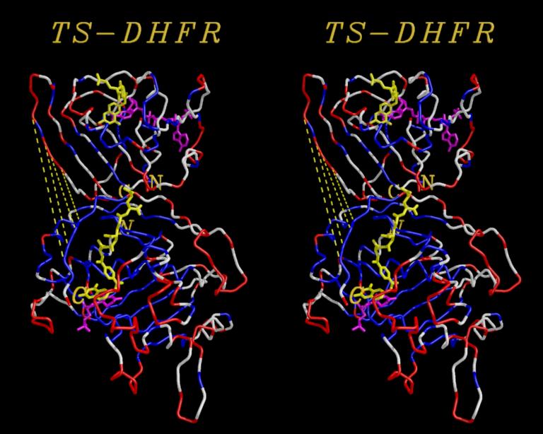
Using the MidasPlus delegates Neon, Label3D and Stereoimg, shown is a cross-eye stereo image of a monomer of E.coli Thymidylate Synthase (lower, TS) and E.coli Dihydrofolate Reductase (upper, DHFR) arranged roughly as determined for TS-DHFR complex (Knighton et. al., Nature Str1994),
The C and N termini of DHFR are labelled in bold, while the C and N termini of TS are labelled in bold italics. Both are colored according to electrostatic potential, where red represents electrostatic values less than -1, white values between -1 and +1, and blue are values greater than +1.
Yellow lines show the pathway along which the DHFR may hinge-bend onto the TS to bring the two protein active sites into close proximity (a theoretical mechanism).
See the manual pages on label3d, stereoimg and neon.