|
|
|
Tom Goddard
April 18, 2019
12:30-3:30 pm
UCSF Parnassus Campus
Medical Sciences Building, room S-163
Organized by Jordan Briscoe Biological Imaging Development Center
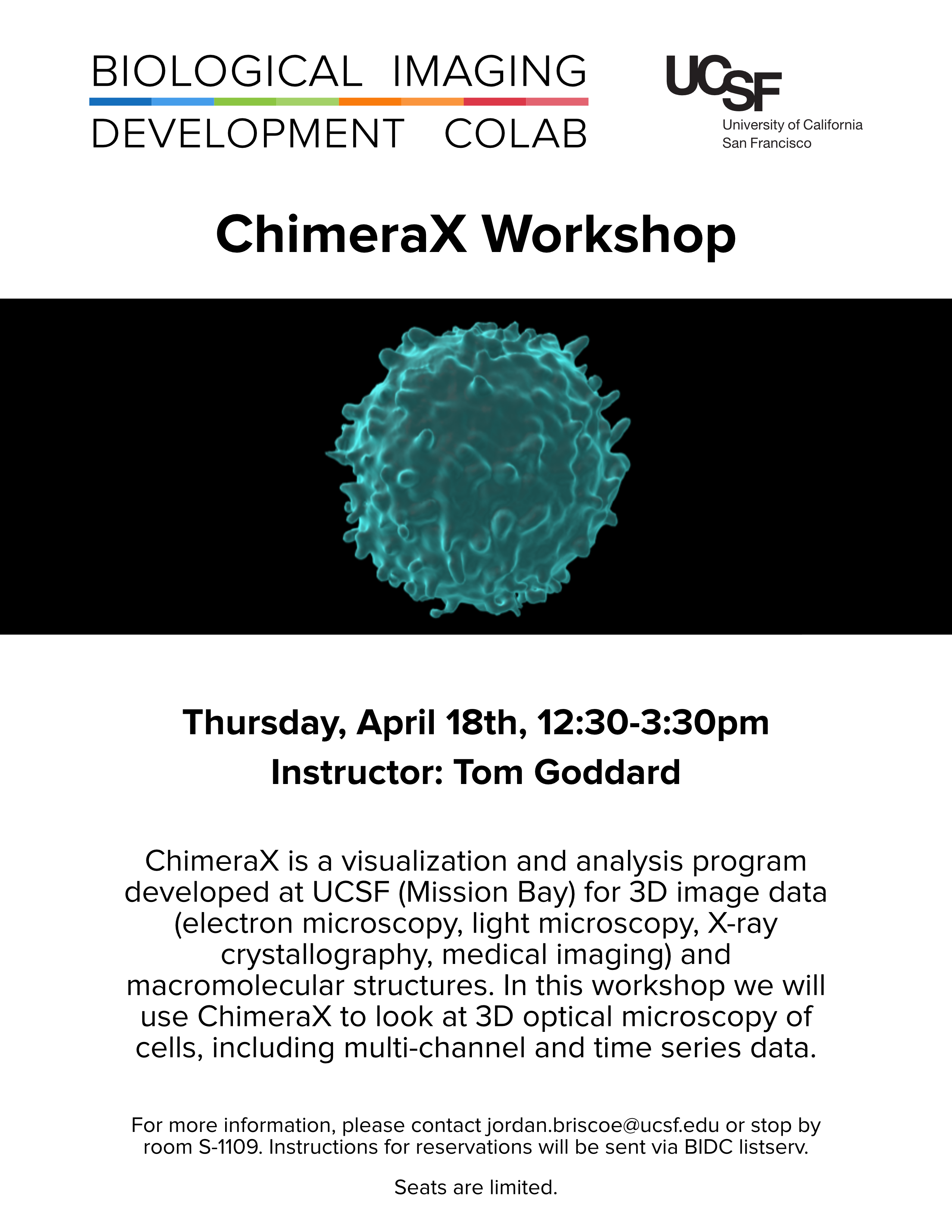
This is a hands-on tutorial for visualization 3D light microscopy data (flyer). Bring your laptop with a ChimeraX daily build installed, and download the tutorial data (250 Mbytes) before the tutorial.
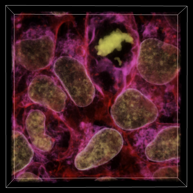
We will look at 3d confocal microscopy of cells with membrane, DNA, and SEC61-beta labeled from the Allen Institute of Cell Science. SEC61-beta is involved in transport of peptides across the endoplasmic reticulum. We will look at different 3D display styles (image, maximum intensity projection, surface, orthogonal planes), brightness, color and transparency control, cropping, masking segmented regions, measuring volumes.
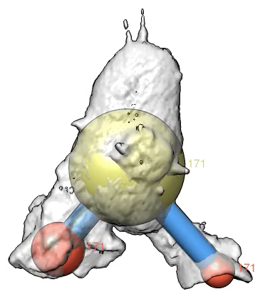
We will also look at a lightsheet microscopy time series of a crawling neutrophil with membrane and actin channels and place markers on the lamellipodia (cell protrusions).