Google™ Search
June 25, 2026
Chimera production release 1.20 is now available,
fixing problems on Mac OS Tahoe
(1.20 release notes).
December 25, 2025
The RBVI wishes you a safe and happy holiday season!
See our
2025 card and the
gallery of previous cards back to 1985.
September 22, 2025
Mac users may wish to defer upgrading to MacOS Tahoe.
Currently on that OS the Chimera graphics window is shifted so that it covers
the command and status lines.
Previous news...
Please note that
UCSF Chimera is legacy software that is no longer being developed or supported.
Users are strongly encouraged to try
UCSF ChimeraX, which is under active development.

UCSF Chimera is a program for the interactive visualization
and analysis of molecular structures and related data,
including density maps, trajectories, and sequence alignments.
It is available free of charge for noncommercial use.
Commercial users, please see
Chimera commercial licensing.
We encourage Chimera users to try ChimeraX
for much better performance with large structures, as well as other major
advantages
and completely new features in addition to nearly all the capabilities
of Chimera (details...).
Chimera is no longer under active development.
Chimera development was supported by a grant from the
National Institutes of Health (P41-GM103311)
that ended in 2018.
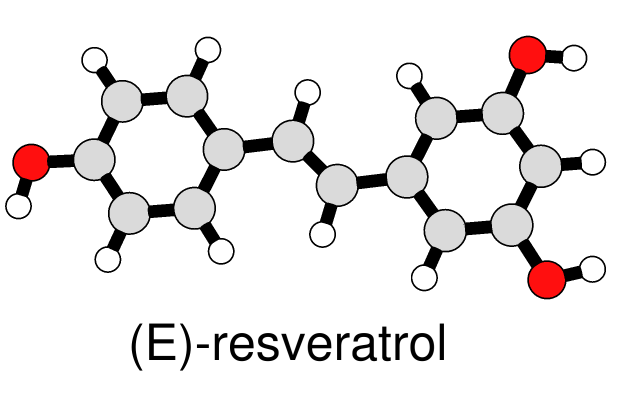
A simple, line-drawing-like appearance can be achieved by combining
black silhouette edges with flat, ambient-only lighting. The image at left
of a small molecule in ball-and-stick style can be generated with a few
Chimera commands.
The same visual effects can be applied to ribbons and other representations,
as shown in
Neuraminidase Flowers
and other Image Gallery entries.
(More features...)
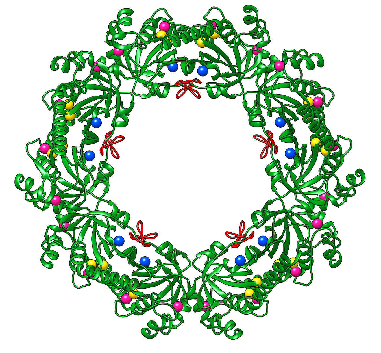
Peroxiredoxins are enzymes that help cells cope with stressors
such as high levels of reactive oxygen species. The image shows a decameric
peroxiredoxin from human red blood cells (Protein Data Bank entry
1qmv),
styled as a holiday wreath.
See also the RBVI
holiday card gallery.
(More samples...)
About RBVI
| Projects
| People
| Publications
| Resources
| Visit Us
Copyright 2018 Regents of the University of California.
All rights reserved.